PLANT COMMUNICATIONS
By Melody Winnig and Vince Giuliano
Plants are far from being deaf and dumb. We think this blog may help convince you that they are smart, listen carefully and are highly expressive.
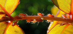
The topic of this blog is plant communication – communications within different parts of one plant, between nearby plants of the same species, between plants of different species they intersect with, between plants and predatory insects that will eat herbivores that are destroying the plant, and multi-way communications between plants and insects and microbial organsms that can help a plant deal with stresses and counter predators. We have much In common with plants, sharing about a quarter of our DNA with them and utilizing many of the biological pathways they use. Besides being fascinating, understanding plants may help us in many ways including how to prevent or limit cancers and other diseases, and how to live long healthy lives. Plant signaling may have strong implications for creating sustainable agriculture, for identifying and understanding health-producing phyto-substances and even for gardening and getting on better with our house plants.  Image source
We first became interested in plant communications after reading well-known biologist Daniel Chamovitz’s book What a Plant Knows. Chamovitz is Director of the Manna Center for Plant Biosciencesat Tel Aviv University). HIs lab is focused primarily on understanding the role of the COP9 signalosome (CSN) in regulating development. The CSN functions at the nexus of signaling inputs and ubiquitin-mediated protein degradation pathways. The lab is using a combination of genetic, molecular and genomic approaches to dissect the varied roles of the CSN in the development of plants and animals, using both Arabidopsis and Drosophila as model systems.
But we will not be getting into such detailed matters here. Readers of this blog know we often love to dig into complex molecular biology pathways. We don’t do that in this blog entry because there are several basics to be told first, and because those pathways in plants are less explored as compared to in animals. Because of the rich variety of plant communications and depth of this field, we necessarily confine ourselves in this introductory blog post to illustrative examples, leaving out much interesting detail.
Why plant communications in a longevity science blog?
Plant signaling is important for humans and for their defenses and longevity. Plant signaling is a defense mechanism for the plants immune system, that is, a system that detects dangers to the wellbeing of an organism and takes defensive actions. Plant communications can warn other parts of the same plant, other plans of the same species, or other nearby plants of different species that there is an invader coming to harm them. This allows a multiplicity of defensive action to be taken. Because plant immunity communications systems work very differently from ours, understanding how they work may facilitate us to augment our own immunity responses and thus contribute to our longevity.
Plants have much in common with us and can exhibit remarkable behavior once thought to be possible only in animals with nervous systems and brains. Plant communications are part of plant behaviors – what plants pro-actively do. These behaviors are varied and complex, despite our perceiving plants just sitting there passively and accepting what comes. Plants behave in respond to light and shade, to the availability of water and nutrients, to heat and cold, to seasons, to what is in their neighborhood, to changes in air composition, to other stresses and to the presence of specific predators. They have memory and predictive capabilities. They can signal to warn themselves of a danger, to warn neighboring plants, to organize individual and collective defenses, and to enroll members of other species that can help them in an emergency. Plants live together in a very complicated and interconnected ecological system. They talk back and forth with many other species. The more we can understand about the communications mechanisms, the more the possibilities for the development of sustainable applications to agricultural and the more we can possibly learn about other species, including ourselves.
The purpose of plant communications, as much as has been established so far by science, is plant development and survival on multiple levels – for individual plants, for communities of plants and for species. It is a defensive mechanism mainly initiated by experienced stresses or survival needs. One important form of signaling we will discuss is release of volatile organic compounds (VOCs), chemical cues given off by a damaged plant. But, as we will see, plants can communicate by counterparts of all of our own sensory modalities.
What this blog entry is not about
There are many forms of molecular plant communications that happen in cell-level and intercellular signaling pathways, just like in bacterial, animal and all other life forms. We mention there is an important class of internal signaling molecules, phytohormones. Phytohormones regulate all the stages of a plants lifecycle and its responses to stresses. A PowerPoint presentation on phytohormones can be downloaded from this site. Two of the phytohormone families are stress responsive and of particular relevance in this regard, Salicylates and Jasmonates Further there is much important internal signaling that takes place via the phloem, an important vascular system in plants. The phloem is made up of live cells – active tubes that transport nutrients, particularly sugars produced by photosynthesis in the leaves. In trees it is the innermost layer of the bark. The phloem as a signaling conduit is covered in the 2005 publication Multitude of Long-Distance Signal Molecules Acting Via Phloem. Also, plants like us humans can utilize microRNAs for inter-cellular and inter organ communications. See “And yet it moves”: Cell-to-cell and long-distance signaling by plant microRNAs. Interesting though these topics are, in this blog entry we will not focus on such basically internal plant signaling pathways. Instead, we focus on forms of communications capable of reaching other plants or organisms belonging to other species.
Plant communication was all the rage in the early 1980’s with the “talking trees” theory popularized in the media. But then the idea was quickly discredited in scientific circles in reaction to several studies that could not be replicated. However, by the late 1990’s a new group of verifiable studies began to emerge re-enforcing the existence of complex signaling process among plants. This blog will present a number of ways that plant signalling/communication/eavesdropping has been documented.
Plants communicate via the same senses humans and animals do, using visual, sound, touch and smell signaling
For communications purposes we may use visual clues, sound, touch, and smell, our communications being based on specialized organs optimized for our survival. Plants too sense and communicate via visual clues, sound, touch and smell – but using completely different perceptive and information processing means. We also communicate internally via electrical signaling, such as at synaptic junctions. Plants also utilize electrical signaling. We provide selected examples of each modality.  Image source
Visual communications
It has been well accepted for some time that Plants reprogram their growth responding to light and shade.Everybody who tends to plants knows this. What they may not know is that plants can predict when shade can be expected from other plants and proactively take actions to keep themselves in the light.
The 1999 publication Keeping up with the neighbours: phytochrome sensing and other signalling mechanisms reported: “Plants ‘forage’ for light in plant canopies using a variety of photosensory systems. Far-red radiation (FR) reflected by neighbours is an early signal of competition that elicits anticipatory shade-avoidance responses. In Arabidopsis and cucumber, perception of reflected FR requires phytochrome B. Horizontal blue (B) light gradients also guide plant shoots to canopy gaps in patchy vegetation, and these B light signals are perceived by specific photoreceptors. When plants are shaded by neighbours they undergo extensive reprogramming of their morphological development. Although phytochromes and B light receptors are certainly involved in these responses to shading, other sensory systems probably play important roles in the field. Recent studies of plant–plant signalling are unveiling a paradigm of sensory diversity and sophistication, which has important implications for understanding the functioning of plant populations and communities.” So, the implicit visual message put out by a plant shooting our new foliage is “Sorry friends, but I have to spread my leaves.” The message received by plants potentially being cast into shade is “Some other plant or plants could be putting me in the shade; I have to do something about that.” The plant response could be long shoots, favoring growth on one side, leaf adaption, etc.
Looking out our office window, we can see semi-shaded patches of wild vegetation, trees, bushes, vines, leafy plants and grasses – sometimes dozens or hundreds of plant species in just a tiny area. We have often wondered how equilibrium could be established involving so many different proximate species. We speculate that this is a result of different species employing somewhat different survival approaches. It all seems very tranquil, but actually there is probably a lot of inter-species arguing, shouting and even screaming going on. The distribution of roots, branches, shoots and leaves is in large measure the collective result of the competitive resource-seeking efforts of all those plants. All that greenery seems to grow naturally and peacefully, but what is happening is an intense multi-way game of anticipatory and responsive communication, competition, and sometimes cooperation in interaction with other species such as fungi and insects. A major resource being light, it seems that trees have a big advantage if and when they can manage to grow up.
Touch communications
It has long been known that simply touching the leaves of some plants can initiate behavioral responses such as leaf curling. If an insect touches two hairs within 20 seconds on the inner surface of a venus flytrap plant, the plant will instantly spring shut entrapping the insect so it can be digested by the plant. There are a great many other examples of “smart” plant sensitivity to touch
The 1990 publication Rain-, wind-, and touch-induced expression of calmodulin and calmodulin-related genes in Arabidopsis reports: “In response to water spray, subirrigation, wind, touch, wounding, or darkness, Arabidopsis regulates the expression of at least four touch-induced (TCH) genes. Ten to thirty minutes after stimulation, mRNA levels increase up to 100-fold. Arabidopsis plants stimulated by touch develop shorter petioles and bolts. This developmental response is known as thigmomorphogenesis. TCH 1 cDNA encodes the putative Arabidopsis calmodulin differing in one amino acid from wheat calmodulin. Sequenced regions of TCH 2 and TCH 3 contain 44% and 70% amino acid identities to calmodulin, respectively. The regulation of this calmodulin-related gene family in Arabidopsis suggests that calcium ions and calmodulin are involved in transduction of signals from the environment, enabling plants to sense and respond to environmental changes.” See also the 1993 publication Thigmomorphogenesis: the effect of mechanical perturbation on plants.
From The Myth of Stoic Trees: “Plants, being immobile, have responses to their environment often quite different from organisms that can escape unsuitable conditions. Therefore, plant responses to touch (often termed mechanical perturbation or MP in the scientific literature) can be exquisitely sensitive. The ability of some carnivorous plants to actively trap food is an example of touch response, as is leaf movement of sensitive plants (Mimosa spp.), and coiling of vine tendrils. == Thigmomorphogenesis can be induced by many types of environmental MP including wind, water spray, snow load, and rubbing from other plants. People, wild and domesticated animals, and even insects can also cause these changes. The responses are species-specific in terms of the amount of MP required and in the morphological changes seen. Initially studied in annual crop plants, such as peas, beans, corn, and sunflowers, MP was universally seen to decrease stem elongation and increase stem thickness. Other characteristics include shorter petiole length, decreased needle elongation, smaller leaves, reduced flower number, and increased senescence (programmed tissue death). Similar responses have been demonstrated in woody species including pine (Pinus), spruce (Picea), fir (Abies), poplar (Populus), and elm (Ulmus). Continual rubbing or brushing of woody trees and shrubs, even that which is gentle enough not to abrade tissue, will result in shorter heights and wider trunks. This is partially meditated through the release of ethylene gas, a naturally produced plant growth regulator, which in turn increases the formation of lignin in the disturbed tissues. The result of thigmomorphogenesis is a stocky, sturdy plant that is more resistant to breakage or windthrow than one that has been untouched, and the greater the disturbance the more pronounced the response. The short, stunted appearance of alpine forest trees is an extreme example of wind-induced thigmomorphogenesis. Such trees are less likely to break from snowload or suffer windthrow than thin, upright specimens.”
So a tree can respond to a massage. But this is not a New Age blog; we are interested in effects documented by hard scientific research.
The 2005 publication In touch: plant responses to mechanical stimuli reports: “Perception and response to mechanical stimuli are likely essential at the cellular and organismal levels. Elaborate and impressive touch responses of plants capture the imagination as such behaviors are unexpected in otherwise often quiescent creatures. Touch responses can turn plants into aggressors against animals, trapping and devouring them, and enable flowers to be active in ensuring crosspollination and shoots to climb to sunlit heights. Morphogenesis is also influenced by mechanical perturbations, including both dynamic environmental stimuli, such as wind, and constant forces, such as gravity. Even individual cells must sense turgor and wall integrity, and subcellular organelles can translocate in response to mechanical perturbations. Signaling molecules and hormones, including intracellular calcium, reactive oxygen species, octadecanoids and ethylene, have been implicated in touch responses. Remarkably, touch-induced gene expression is widespread; more than 2.5% of Arabidopsis genes are rapidly up-regulated in touch-stimulated plants. Many of these genes encode calcium-binding, cell wall modifying, defense, transcription factor and kinase proteins. With these genes as tools, molecular genetic methods may enable elucidation of mechanisms of touch perception, signal transduction and response regulation.”
The 2012 publication Arabidopsis touch-induced morphogenesis is jasmonate mediated and protects against pests reports: “Plants cannot change location to escape stressful environments. Therefore, plants evolved to respond and acclimate to diverse stimuli, including the seemingly innocuous touch stimulus [1-4]. Although some species, such as Venus flytrap, have fast touch responses, most plants display more gradual touch-induced morphological alterations, called thigmomorphogenesis [2, 3, 5, 6]. Thigmomorphogenesis may be adaptive; trees subjected to winds develop less elongated and thicker trunks and thus are less likely damaged by powerful wind gusts [7]. Despite the widespread relevance of thigmomorphogenesis, the regulation that underlies plant mechanostimulus-induced morphological responses remains largely unknown. Furthermore, whether thigmomorphogenesis confers additional advantage is not fully understood. — Here we show that jasmonate (JA) phytohormone both is required for and promotes the salient characteristics of thigmomorphogenesis in Arabidopsis, including a touch-induced delay in flowering and rosette diameter reduction. Furthermore, we find that repetitive mechanostimulation enhances Arabidopsis pest resistance in a JA-dependent manner. These results highlight an important role for JA in mediating mechanostimulus-induced plant developmental responses and resultant cross-protection against biotic stress.”
Smell communications
A central focus here is on molecular be communications via VOCs (volatile organic compounds) – giving off odors and smelling in animal terms. This is an area where plants do a better job than humans based on their millions of years of evolution, a very essential communications means for plants. These VOCs can inform other parts of a plant or other nearby plants to respond/emit chemicals that ward off attackers/curl leaves etc to repel the attacker. The distance VOCs travel, for the most part is 60cm or less though some only travel 15cm. It is not so well established if the intention is only to inform nearby plants of the same species or not. But there is often a defense response by other plant species if they are close enough. First, we will frame this branch of plant science research in a historical context.
The double about-face of science about plant smell communications
There were publications in the 1980s that plants communicate via volatile substances with each other about threats of being eaten. These publications were flawed in their research methodology and were soon discredited by publications from reputable scientists. However, evidence for such effects continued to accumulate and now there is little doubt that such communication exists. Plants of a number of species receiving communications from plants threatened or under attack mount defenses and are less likely to be eaten.
The 2014 publication Volatile communication between plants that affects herbivory: a meta-analysis Ecology Letters reviews the situation with respect to VOC signaling caused by herbivore attacks, i.e. signaling from the plant when it is being eaten or threatened to be eaten. The publication states in summary “Volatile communication between plants causing enhanced defense has been controversial. Early studies were not replicated, and influential reviews questioned the validity of the phenomenon. We collected 48 well-replicated studies and found overall support for the hypothesis that resistance increased for individuals with damaged neighbours. Laboratory or greenhouse studies and those conducted on agricultural crops showed stronger induced resistance than field studies on undomesticated species, presumably because other variation had been reduced. A cumulative analysis revealed that early, non-replicated studies were more variable and showed less evidence for communication. — These results indicate that plants of diverse taxonomic affinities and ecological conditions become more resistant to herbivores when exposed to volatiles from damaged neighbours.”
First statements of the volatile communications hypothesis
Going on: “When some plants are attacked by herbivores, they release chemical cues that cause other individuals to change their traits and become more resistant to herbivory. Communication between plants was first observed and reported more than 30 years ago (Rhoades 1983; Baldwin & Schultz 1983) and the number of reported cases has grown rapidly in the recent past (Heil & Karban 2010). We consider a process to be plant volatile communication if it involves signalling by a plant that causes a response in the same or a different individual that receives the cue. Emission or display of a cue is plastic and the response of the receiver is conditional on receiving the cue. We require that emitting the cue could potentially benefit the emitter, although this has proven difficult or impossible to establish (Karban 2008). For an interaction to be considered communication, the responder must have the choice of responding to the cue or not, a requirement which excludes allelopathy (Schenk & Seabloom 2010). We make no assumptions about the intended target of the cues; many plants use volatile cues to co-ordinate their own defences against herbivores when one branch is attacked and other branches on the same individual respond by increasing defences (Karban et al. 2006; Frost et al. 2007; Heil & Silva Bueno 2007; Rodriguez-Saona et al. 2009). There is no agreed upon definition of communication; our use of the term is broader than most and includes phenomena that some authors prefer to call eavesdropping or signalling. — Early reports of plant communication met with great interest from scientists and the popular press. David Rhoades observed that caterpillars placed on willow trees near damaged neighbours grew less well than those placed on trees near undamaged neighbours (Rhoades 1983). He hypothesised that the reduction in performance was caused by airborne communication from the damaged trees that increased resistance in neighbours. However, he was unable to repeat his initial results and his experimental design lacked true replication (D. Rhoades, pers. comm., Fowler & Lawton 1985). In addition, the poor performance of caterpillars on trees in close proximity to infested neighbours could have been caused by the introduction of insect pathogens rather than by communication between plants. Early experiments conducted on plants in growth chambers reported that plants exposed to volatiles coming from chambers containing feeding herbivores became more resistant, but the experimental designs used in these studies also lacked true replication (Baldwin & Schultz 1983; Bruin et al. 1992).”
The first about-face
“Following an influential review by Simon Fowler and John Lawton, most ecologists decided that communication between plants was a phenomenon that had been considered and debunked and that the phenomenon did not occur in nature (Fowler & Lawton 1985; Dicke & Bruin 2001). In addition to the limitations associated with experimental design of the early studies, communication between plants that benefited neighbours did not make sense to many ecologists. Early descriptions of this phenomenon were referred to as ‘talking trees’ by both the popular press and some scientists in the field. Natural selection would not be expected to favour the emission of cues that provided neighbouring competitors with information about herbivores.”
The second about-face
“Coinciding with the resurgence in scientific interest in communication has been a resolution of this apparent contradiction. — Many studies involving diverse plants reported evidence of volatile communication resulting in increased resistance to herbivore attack, indicating that this is a widespread natural phenomenon. Unlike early studies of this phenomenon, the studies considered in this review were well-replicated with independent sampling units. — Many of these studies did not identify mechanisms involved, even whether a plant response was responsible for the effects. Alternative hypotheses, such as direct repellency of herbivores by volatiles, were often not ruled out and would be worth considering in future studies. Determining the plant responses involved, particularly the volatile cues that were responsible will be well worth future effort. As expected, conditions that minimised background variation, particularly laboratory studies and studies of genetically homogeneous crop species, were more likely to detect significant effects of volatile cues on induced resistance. Future studies are needed to separate effects due to experimental conditions (laboratory vs. field) from those caused by different inducing agents (herbivores vs. artificial damage”
Up to this point in our discussion: 1. We know that communications among plants warning of herbivore attacks must exist because plants that are neighbors of attacked plants are less damaged in subsequent attacks, 2. With high probability VOCs are involved, 3. The exact mechanisms of initiation of communications, forms of protection and mechanisms of protection are less studied. We go on to discuss these issues.
Plant VOC communications in response to predatory attacks can be highly specific and can involve multiple kinds of VOCs including release of 1. substances that are repellant to the specific predator, 2. substances that are toxic to the specific predator, 3 substances that are alerts to other parts of the same plant or neighboring plants that tell them to upgrade a kind of predator defense, and 4. substances that attract natural enemies of a predator.
VOC interspecies communications that attract natural enemies of predators
There are many examples of this phenomenon. The 2011 publication A cry for help from leaf to root: Above ground insect feeding leads to the recruitment of rhizosphere microbes for plant self-protection against subsequent diverse attacks relates “Plants have evolved general and specific defense mechanisms to protect themselves from diverse enemies, including herbivores and pathogens. To maintain fitness in the presence of enemies, plant defense mechanisms are aimed at inducing systemic resistance: in response to the attack of pathogens or herbivores, plants initiate extensive changes in gene expression to activate “systemic acquired resistance” against pathogens and “indirect defense” against herbivores. Recent work revealed that leaf infestation by whiteflies, stimulated systemic defenses against both an airborne pathogen and a soil-borne pathogen, which was confirmed by the detection of the systemic expression of pathogenesis-related genes in response to salicylic acid and jasmonic acid-signaling pathway activation. Further investigation revealed that plants use self protection mechanisms against subsequent herbivore attacks by recruiting beneficial microorganisms called plant growth-promoting rhizobacteria/fungi, which are capable of reducing whitefly populations. Our results provide new evidence that plant-mediated aboveground to belowground communication and vice versa are more common than expected.”
An additional important point made here is that different predator and pathogen attacks can activate the same signaling pathway that upgrades complexes of defensive genes. The result is strengthened resistance to a variety of future attacks.
This is similar to hormesis as expressed on animals, a topic much-discussed in this blog. Going on with examples of VOC communications:
It has not been determined what plants are the intended target of the volatile signaling cues. It is possible that main targets are different branches of the same plant which are not sufficiently connected vascularly. However, the following several research papers show different ways plants can use volatiles to stimulate their own defenses against herbivores.
The case of hybrid poplar vs gypsy moth larvae shows how when one branch is attacked, other branches on the same plant respond by increasing their defenses
The 2007 publication Within-plant signalling via volatiles overcomes vascular constraints on systemic signalling and primes responses against herbivores makes this point: “Plant volatiles play important roles in signaling between plants and insects, but their role in communication among plants remains controversial. Previous research on plant–plant communication has focused on interactions between neighbouring plants, largely overlooking the possibility that volatiles function as signals within plants. Here, we show that volatiles released by herbivore-wounded leaves of hybrid poplar (Populus deltoides × nigra) prime defences in adjacent leaves with little or no vascular connection to the wounded leaves. Undamaged leaves exposed to volatiles from wounded leaves on the same stem had elevated defensive responses to feeding by gypsy moth larvae (Lymantria dispar L.) compared with leaves that did not receive volatiles. Volatile signals may facilitate systemic responses to localized herbivory even when the transmission of internal signals is constrained by vascular connectivity. Self-signalling via volatiles is consistent with the short distances over which plant response to airborne cues has been observed to occur and has apparent benefits for emitting plants, suggesting that within-plant signalling may have equal or greater ecological significance than signalling between plants.”
Volatile signaling can work within the plant sagebrush, (Artemisia tridentata) and between plants up to 60 cm apart
The 2006 publication Damage-induced resistance in sagebrush: volatiles are key to intra- and interplant communication reports “”Airborne communication between individuals, called “eavesdropping” in this paper, can cause plants to become more resistant to herbivores when a neighbor has been experimentally clipped. The ecological relevance of this result has been in question, since individuals may be too far apart for this interaction to affect many plants in natural populations. We investigated induced resistance to herbivory in sagebrush, Artemisia tridentata, caused by experimental clipping of the focal plant and its neighbors. We found no evidence for systemic induced resistance when one branch was clipped and another branch on the same plant was assayed for naturally occurring damage. In this experiment, air contact and plant age were not controlled. Previous work indicated that sagebrush received less damage when a neighboring upwind plant within 15 cm had been experimentally clipped. Here we found that pairs of sagebrush plants that were up to 60 cm apart were influenced by experimental clipping of a neighbor. Furthermore, we observed that most individuals had conspecific neighbors that were much closer than 60 cm. Air contact was essential for communication; treatments that reduced airflow between neighboring individuals, either because of wind direction or bagging, prevented induced resistance. Airflow was also necessary for systemic induced resistance among branches within an individual. Reports from the literature indicated that sagebrush is highly sectorial, as are many desert shrubs. Branches within a sagebrush plant do not freely exchange material via vascular connections and apparently cannot rely on an internal signaling pathway for coordinating induction of resistance to herbivores. Instead, they may use external, volatile cues. This hypothesis provides a proximal explanation for why sagebrush does not demonstrate systemic induced resistance without directed airflow, and why airborne communication between branches induces resistance.”
When lima bean plants are attacked by herbivores, the plants give off volatiles thatattract natural predators of the herbivores, not only affecting the attacked plants but also the neighboring plants. But this does not happen when they are exposed to artificially wounded plants.
Lima beans vs. spider mites; mobilizing spider mites
The 2000 publication Herbivory-induced volatiles elicit defence genes in lima bean leaves is one of a number of relevant publications on plant VOC communications that started to occur in the early 2000s and that highlight the interspecies nature of the communications: “In response to herbivore damage, several plant species emit volatiles that attract natural predators of the attacking herbivores1, 2, 3, 4, 5. Using spider mites (Tetranychus urticae) and predatory mites (Phytoseiulus persimilis)1, 2, 3, 4, it has been shown that not only the attacked plant but also neighbouring plants are affected, becoming more attractive to predatory mites 3, 6 and less susceptible to spider mites 6. The mechanism involved in such interactions, however, remains elusive. Here we show that uninfested lima bean leaves activate five separate defence genes when exposed to volatiles from conspecific leaves infested with T. urticae, but not when exposed to volatiles from artificially wounded leaves. The expression pattern of these genes is similar to that produced by exposure to jasmonic acid. At least three terpenoids in the volatiles are responsible for this gene activation; they are released in response to herbivory but not artificial wounding. Expression of these genes requires calcium influx and protein phosphorylation/dephosphorylation.”
Lima beans vs. beetles; mobilizing predatory anthropods
A subsequent publication on lima bean plants appearing in 2007 was Within-plant signaling by volatiles leads to induction and priming of an indirect plant defense in nature: “Plants respond to herbivore attack with the release of volatile organic compounds (VOCs), which can attract predatory arthropods and/or repel herbivores and thus serve as a means of defense against herbivores. Such VOCs might also be perceived by neighboring plants to adjust their defensive phenotype according to the present risk of attack. We exposed lima bean plants at their natural growing site to volatiles of beetle-damaged conspecific shoots. This reduced herbivore damage and increased the growth rate of the exposed plants. To investigate whether VOCs also can serve in signaling processes within the same individual plant we focused on undamaged “receiver” leaves that were either exposed or not exposed to VOCs released by induced “emitter” leaves. Extrafloral nectar secretion by receiver leaves increased when they were exposed to VOCs of induced emitters of neighboring plants or of the same shoot, yet not when VOCs were removed from the system. Extrafloral nectar attracts predatory arthropods and represents an induced defense mechanism. The volatiles also primed extrafloral nectar secretion to show an augmented response to subsequent damage. Herbivore-induced VOCs elicit a defensive response in undamaged plants (or parts of plants) under natural conditions, and they function as external signal for within-plant communication, thus serving also a physiological role in the systemic response of a plant to local damage.”
When alders are attacked by the alder leaf beetle, their undamaged neighbor alders also respond with increased resistance to the alder beetle leaf herbivory. The lab experiments also supported field results. Defoliation of alders may trigger interplant resistance transfer, and therefore reduce herbivory in whole alder stands.
Alders vs. alder leaf beetles
Another 2000 publication that helped facilitate the swing back to acceptance of plant VOC communications was Defoliation of alders (Alnus glutinosa) affects herbivory by leaf beetles on undamaged neighbors: “The effects of defoliation of alder (Alnus glutinosa) on subsequent herbivory by alder leaf beetle (Agelastica alni) were studied in ten alder stands in northern Germany. At each site, one tree was manually defoliated (c.20% of total foliage) to simulate herbivory. Subsequent damage by A. alniwas assessed on ten alders at each site on six different dates from May to September 1994. After defoliation, herbivory by A. alni increased with distance from the defoliated tree. Laboratory experiments supported the field results. Not only leaf damage in the field, but also the extent of leaf consumption in laboratory feeding-preference tests and the number of eggs oviposited per leaf in another laboratory test were positively correlated with distance from the defoliated tree. Resistance was therefore induced not only in defoliated alders, but also in their undamaged neighbours. Consequently, defoliation of alders may trigger interplant resistance transfer, and therefore reduce herbivory in whole alder stands.”
Sound communications
Plants can also respond to vibrational communications. The 2014 publication Plants respond to leaf vibrations caused by insect herbivore chewing reports: “Plant germination and growth can be influenced by sound, but the ecological significance of these responses is unclear. We asked whether acoustic energy generated by the feeding of insect herbivores was detected by plants. We report that the vibrations caused by insect feeding can elicit chemical defenses. Arabidopsis thaliana (L.) rosettes pre-treated with the vibrations caused by caterpillar feeding had higher levels of glucosinolate and anthocyanin defenses when subsequently fed upon by Pieris rapae (L.) caterpillars than did untreated plants. The plants also discriminated between the vibrations caused by chewing and those caused by wind or insect song. Plants thus respond to herbivore-generated vibrations in a selective and ecologically meaningful way. A vibration signaling pathway would complement the known signaling pathways that rely on volatile, electrical, or phloem-borne signals. We suggest that vibration may represent a new long distance signaling mechanism in plant–insect interactions that contributes to systemic induction of chemical defenses.”
Continuing: “The effects of sound on plant growth and other traits have been recognized for decades, but the ecological significance of these responses is unclear. While plant responses to wind and touch have been examined and have clear adaptive significance (Chehab et al. 2009), plant responses to acoustic energy have largely been studied in the absence of an ecological context. For example, there is a long tradition of exposing plants to musical sound (Klein and Edsall 1965; Telewski 2006; Jeong et al. 2004). Although music influences growth and germination in some plants, music contains such a wide range of frequencies, amplitudes and fine-temporal patterns that its usefulness as an experimental stimulus is limited. More systematic studies have found that some frequencies have a greater influence than others (Telewski 2006). For example, young roots of corn grow towards the source of continuous tones, transmitted as airborne or waterborne sound, and respond optimally to frequencies of 200–300 Hz (Gagliano et al. 2012a). While these studies bring us a step closer to being able to link plant responses to acoustic energy to ecologically relevant sound sources, the experimental stimuli still remain far removed from those produced by natural sources of acoustic energy in the plant’s environment. — One of the most relevant sources of acoustic energy in the immediate environment of a plant is the rich community of plant-associated arthropods, including herbivores, predators, and parasitoids (Cocroft and Rodriguez 2005). Plant-borne vibrations provide a wealth of information about the activities of insects on plants. Within the abundant arthropod community on plants, many ecological and social interactions depend on the perception and production of plant-borne vibrations (Hill 2008). Some 200,000 species of insects communicate using substrate vibrations to locate mates, attract mutualists, or exploit plant resources (Cocroft and Rodriguez 2005). Many more insects and other arthropods use such vibrations to locate prey or avoid predators (Barth 1998; Castellanos and Barbosa 2006; Casas and Magal 2006; Virant-Doberlet et al.2011; Cocroft 2011). Chewing herbivores, in particular, produce characteristic, high-amplitude vibrations that travel rapidly to other parts of the plant. Predatory insects can use chewing vibrations to detect their prey from a considerable distance: for example, on soybean, the chewing vibrations of green clover worms elicited search by predatory stinkbugs 50 cm away (Pfannenstiel et al. 1995). We suggest that the vibrations produced by chewing herbivores are an important source of acoustic energy for plants. If plants can detect and use this conspicuous, reliable and rapidly transmitted source of information about herbivore feeding, tissues far from the site of attack could use feeding vibrations to respond quickly to the threat of herbivory. A vibration signaling pathway would complement the known signaling pathways that rely on phloem-borne signals, airborne volatiles, or electrical signals (Wu and Baldwin 2009; Mousavi et al. 2013). Here we test the hypothesis that plant responses to herbivory, in the form of induced chemical defenses, can be elicited by the mechanical vibrations produced by chewing caterpillars. We report that Arabidopsis thaliana plants exposed to chewing vibrations produced greater amounts of chemical defenses in response to subsequent herbivory, and that the plants distinguished chewing vibrations from other environmental vibrations.”
Root communications
This is a form of extended communications we humans do not have.
Touch and chemical communications can also occur via undergrownd root-connecting mycelial networks, e,g, networks of fungal filaments. The 2012 publication Mycorrhiza-induced resistance and priming of plant defenses reports: “Symbioses between plants and beneficial soil microorganisms like arbuscular-mycorrhizal fungi (AMF) are known to promote plant growth and help plants to cope with biotic and abiotic stresses. Profound physiological changes take place in the host plant upon root colonization by AMF affecting the interactions with a wide range of organisms below- and above-ground. Protective effects of the symbiosis against pathogens, pests, and parasitic plants have been described for many plant species, including agriculturally important crop varieties. Besides mechanisms such as improved plant nutrition and competition, experimental evidence supports a major role of plant defenses in the observed protection. During mycorrhiza establishment, modulation of plant defense responses occurs thus achieving a functional symbiosis. As a consequence of this modulation, a mild, but effective activation of the plant immune responses seems to occur, not only locally but also systemically. This activation leads to a primed state of the plant that allows a more efficient activation of defense mechanisms in response to attack by potential enemies. Here, we give an overview of the impact on interactions between mycorrhizal plants and pathogens, herbivores, and parasitic plants, and we summarize the current knowledge of the underlying mechanisms. We focus on the priming of jasmonate-regulated plant defense mechanisms that play a central role in the induction of resistance by arbuscular mycorrhizas.”
Linking into a mycelia network can have a profound positive effect on gene expression health state and disease resistance in the linked plants.
The 2007 publication Arbuscular mycorrhizal symbiosis is accompanied by local and systemic alterations in gene expression and an increase in disease resistance in the shoots reports: “In natural ecosystems, the roots of many plants exist in association with arbuscular mycorrhizal (AM) fungi, and the resulting symbiosis has profound effects on the plant. The most frequently documented response is an increase in phosphorus nutrition; however, other effects have been noted, including increased resistance to abiotic and biotic stresses. Here we used a 16,000-feature oligonucleotide array and real-time quantitative RT-PCR to explore transcriptional changes triggered in Medicago truncatula roots and shoots as a result of AM symbiosis. By controlling the experimental conditions, phosphorus-related effects were minimized, and both local and systemic transcriptional responses to the AM fungus were revealed. The transcriptional response of the roots and shoots differed in both the magnitude of gene induction and the predicted functional categories of the mycorrhiza-regulated genes. In the roots, genes regulated in response to three different AM fungi were identified, and, through split-root experiments, an additional layer of regulation, in the colonized or non-colonized sections of the mycorrhizal root system, was uncovered. Transcript profiles of the shoots of mycorrhizal plants indicated the systemic induction of many genes predicted to be involved in stress or defense responses, and suggested that mycorrhizal plants might display enhanced disease resistance. Experimental evidence supports this prediction, and mycorrhizal M. truncatula plants showed increased resistance to a virulent bacterial pathogen, Xanthomonas campestris. Thus, the symbiosis is accompanied by a complex pattern of local and systemic changes in gene expression, including the induction of a functional defense response.”
Electrical communications
Animals use electrical impulses for internal communications, for example at nerve synaptic junctions in muscle cells. Plants also use internal electrical signaling in stress situations, as illustrated in the 2014 publication Real-time, in vivo intracellular recordings of caterpillar-induced depolarization waves in sieve elements using aphid electrodes, Electrical communications are transmitted via the pholem.: “Summary:
- Plants propagate electrical signals in response to artificial wounding. However, little is known about the electrophysiological responses of the phloem to wounding, and whether natural damaging stimuli induce propagating electrical signals in this tissue.
- Here, we used living aphids and the direct current (DC) version of the electrical penetration graph (EPG) to detect changes in the membrane potential of Arabidopsis sieve elements (SEs) during caterpillar wounding.
- Feeding wounds in the lamina induced fast depolarization waves in the affected leaf, rising to maximum amplitude (c. 60 mV) within 2s. Major damage to the midvein induced fast and slow depolarization waves in unwounded neighbor leaves, but only slow depolarization waves in non-neighbor leaves. The slow depolarization waves rose to maximum amplitude (c. 30 mV) within 14 s. Expression of a jasmonate-responsive gene was detected in leaves in which SEs displayed fast depolarization waves. No electrical signals were detected in SEs of unwounded neighbor leaves of plants with suppressed expression of GLR3.3 and GLR3.6.
- EPG applied as a novel approach to plant electrophysiology allows cell-specific, robust, real-time monitoring of early electrophysiological responses in plant cells to damage, and is potentially applicable to a broad range of plant–herbivore interactions”
Multimodal communications involving mixed communications means between species both above and below ground
- Now, we provide examples of interspecies plant communications of increasing complexity.
Underground mycelia networks can provide early warning between plants of an impending aphid attack.
Bean plants vs. aphids; mobilizing parasitoids using a fungal communications channel
The 2013 publication Underground signals carried through common mycelial networks warn neighbouring plants of aphid attack reports: “The roots of most land plants are colonised by mycorrhizal fungi that provide mineral nutrients in exchange for carbon. Here, we show that mycorrhizal mycelia can also act as a conduit for signalling between plants, acting as an early warning system for herbivore attack. Insect herbivory causes systemic changes in the production of plant volatiles, particularly methyl salicylate, making bean plants, Vicia faba, repellent to aphids but attractive to aphid enemies such as parasitoids. We demonstrate that these effects can also occur in aphid-free plants but only when they are connected to aphid-infested plants via a common mycorrhizal mycelial network. This underground messaging system allows neighbouring plants to invoke herbivore defences before attack. Our findings demonstrate that common mycorrhizal mycelial networks can determine the outcome of multitrophic interactions by communicating information on herbivore attack between plants, thereby influencing the behaviour of both herbivores and their natural enemies.” We have at least four different species involved in this case.
Further complex plant communications and responses can lead to complex responses, such as a below ground insect attack leading to above ground protection against attack by insects of a different species.
The 2008 publication Plants as green phones: Novel insights into plant-mediated communication between below- and above-ground insects reports “Plants can act as vertical communication channels or ‘green phones’ linking soil-dwelling insects and insects in the aboveground ecosystem. When root-feeding insects attack a plant, the direct defense system of the shoot is activated, leading to an accumulation of phytotoxins in the leaves. The protection of the plant shoot elicited by root damage can impair the survival, growth and development of aboveground insect herbivores, thereby creating plant-based functional links between soil-dwelling insects and insects that develop in the aboveground ecosystem. The interactions between spatially separated insects below- and aboveground are not restricted to root and foliar plant-feeding insects, but can be extended to higher trophic levels such as insect parasitoids. Here we discuss some implications of plants acting as communication channels or ‘green phones’ between root and foliar-feeding insects and their parasitoids, focusing on recent findings that plants attacked by root-feeding insects are significantly less attractive for the parasitoids of foliar-feeding insects.”
Interaction communications among a plant, fungi, insects and other plants can be complex, multimodal and multidirectional
In the real world it gets a lot more complicated. The 2013 publication Two-way plant mediated interactions between root-associated microbes and insects: from ecology to mechanisms reports: “Plants are members of complex communities and function as a link between above- and below-ground organisms. Associations between plants and soil-borne microbes commonly occur and have often been found beneficial for plant fitness. Root-associated microbes may trigger physiological changes in the host plant that influence interactions between plants and aboveground insects at several trophic levels. Aboveground, plants are under continuous attack by insect herbivores and mount multiple responses that also have systemic effects on belowground microbes. Until recently, both ecological and mechanistic studies have mostly focused on exploring these below- and above-ground interactions using simplified systems involving both single microbe and herbivore species, which is far from the naturally occurring interactions. Increasing the complexity of the systems studied is required to increase our understanding of microbe–plant–insect interactions and to gain more benefit from the use of non-pathogenic microbes in agriculture. In this review, we explore how colonization by either single non-pathogenic microbe species or a community of such microbes belowground affects plant growth and defense and how this affects the interactions of plants with aboveground insects at different trophic levels. Moreover, we review how plant responses to foliar herbivory by insects belonging to different feeding guilds affect interactions of plants with non-pathogenic soil-borne microbes. The role of phytohormones in coordinating plant growth, plant defenses against foliar herbivores while simultaneously establishing associations with non-pathogenic soil microbes is discussed.”
There is a growing interest in all the complex interactions of plants with other plants and organisms and how this affects ecological systems.
The 2012 publication Root herbivore effects on aboveground multitrophic interactions: patterns, processes and mechanisms reports: “In terrestrial food webs, the study of multitrophic interactions traditionally has focused on organisms that share a common domain, mainly above ground. In the last two decades, it has become clear that to further understand multitrophic interactions, the barrier between the belowground and aboveground domains has to be crossed. Belowground organisms that are intimately associated with the roots of terrestrial plants can influence the levels of primary and secondary chemistry and biomass of aboveground plant parts. These changes, in turn, influence the growth, development, and survival of aboveground insect herbivores. The discovery that soil organisms, which are usually out of sight and out of mind, can affect plant-herbivore interactions aboveground raised the question if and how higher trophic level organisms, such as carnivores, could be influenced. At present, the study of above-belowground interactions is evolving from interactions between organisms directly associated with the plant roots and shoots (e.g., root feeders – plant – foliar herbivores) to interactions involving members of higher trophic levels (e.g., parasitoids), as well as non-herbivorous organisms (e.g., decomposers, symbiotic plant mutualists, and pollinators). This multitrophic approach linking above- and belowground food webs aims at addressing interactions between plants, herbivores, and carnivores in a more realistic community setting. The ultimate goal is to understand the ecology and evolution of species in communities and, ultimately how community interactions contribute to the functioning of terrestrial ecosystems. Here, we summarize studies on the effects of root feeders on aboveground insect herbivores and parasitoids and discuss if there are common trends. We discuss the mechanisms that have been reported to mediate these effects, from changes in concentrations of plant nutritional quality and secondary chemistry to defense signaling. Finally, we discuss how the traditional framework of fixed paired combinations of root- and shoot-related organisms feeding on a common plant can be transformed into a more dynamic and realistic framework that incorporates community variation in species, densities, space and time, in order to gain further insight in this exciting and rapidly developing field.”
Plants, predators and bacteria can form various alliances for survival. In some cases the plants come out ahead, in other cases they do not.
An example of the second kind of situation is described in a 2013 Science Daily article Microbes help beetles defeat plant defenses: “Some symbiotic bacteria living inside Colorado potato beetles can trick plants into reacting to a microbial attack rather than that of a chewing herbivore, according to a team of Penn State researchers who found that the beetles with bacteria were healthier and grew better. “For the last couple of decades, my lab has focused on induced defenses in plants,” said Gary W. Felton, professor and head of entomology. “We had some clues that oral secretions of beetles suppressed defenses, but no one had followed up on that research.” Seung Ho Chung, graduate student in entomology working with Felton, decided to investigate how plants identified chewers and how herbivores subverted the plants defenses. “I thought we could identify what was turning the anti-herbivore reaction off,” said Felton. “But it was a lot more difficult because we had not considered microbes.” According to Felton, the beetles do not have salivary glands and so they regurgitate oral secretions onto the leaves to begin digestion. These secretions contain gut bacteria. — Plant defenses against chewing insects follow a jasmonite-mediated pathway that induces protease inhibitors and polyphenol oxidase, which suppress digestion and growth. Plant defenses against pathogens follow a salicylic acid mediated pathway. When the antimicrobial response turns on, it interferes with the response to chewing, allowing the beetles to develop more normally.”
“Chung and Felton used tomato plants to identify exactly what was turning off the response to chewing. They note, however, that the Colorado potato beetle also attacks eggplant and potato plants. They report the results of their work in the current online edition of the Proceedings of the National Academy of Sciences. — The researchers allowed beetle larva to feast on antibiotic-treated leaves and natural leaves and found that on the antibiotic-treated leaves, the beetles suffered from the plant’s anti-herbivore defense, but on the natural leaves the larva gained more weight and thrived. Chung and Felton then investigated expression of genes in the anti-herbivore pathway and the production of enzymes. They found that the presence of bacteria decreases the anti-herbivore response. — The researchers also isolated and grew the bacteria from the Colorado potato beetle guts. They found 22 different types of bacteria, but only three types suppressed the anti-herbivore response. During a variety of other experiments, they found that in all cases presence of the bacteria that could suppress the anti-herbivore response led to healthier beetles.”
Chemical communication from a plant to members of another species can be designed to create an addiction which enforces a long-term symbiotic relationship between the plant and members of that species
Phony messaging is one dirty trick that can be used in nature. Another is creating an addiction. An example is that a plant can create an addiction that keeps members of another species supporting the plant. One such case is explained in a 2013 National Geographic article Trees Trap Ants Into Sweet Servitude “An evolutionary alliance between trees and the ants that guard them has a sinister explanation, a new study suggests, finding ants hooked on nectar. In Central America, ants act as bodyguards for acacia trees, defending them from weeds and hungry animals in exchange for room and board, one of the most iconic alliances in nature. (See “Ant Bodyguards Get Exclusive Contract From Trees.”) But Martin Heil of Cinvestav Unidad Irapuato in Mexico has found that the tree’s sugary snacks are laced with an enzyme that prevents the ants from eating other sources of sugar. One sip, and the insects are consigned to a life of indentured servitude. “It was surprising to me that the immobile, ‘passive’ plant can manipulate the seemingly much more active partner, the ant,” says Heil. The report illustrates how evolution keeps cooperative relationships among some species going, even when one partner is clearly reaping most of the benefits in the arrangement. (See “Video—Acacia Tree Ants.”)”
“Nectar Addicts:Heil compares the tree to a dairy company that sells lactose-free milk that has been chemically altered to render its customers unable to digest normal milk. Any customer who drank it would be forced to stick to that one lactose-free brand. Here’s how it works. Most of the sweet foods that ants eat, such as plant sap, are rich in a sugar called sucrose. The ants digest this with an enzyme called invertase, which breaks sucrose into smaller sugars. In 2005, Heil showed that all of the workers of the acacia ant Pseudomyrmex ferrugineus lack invertase activity and cannot digest normal sources of sucrose. Fortunately, the tree compensates for this impairment by secreting invertase into its nectar, providing the ants with a predigested meal. For this reason, the ants prefer acacia nectar over other sugar sources. But Heil suspected that this explanation was too neat. Why would the ants so heavily restrict their dietary options by losing an important enzyme? Weirder still, in 2008, Heil showed that the larvae have invertase, which becomes deactivated only when they turn into adults. Culprit Nabbed: Heil has now shown that the tree itself is responsible. Writing in the Ecology Letters journal, he reports that acacia nectar contains chitinase enzymes that completely block invertase. Shortly after the workers emerge from their pupae as adults, they take their first sip of nectar and their invertase becomes irreversibly disabled. “Ain’t nature grand?” says Todd Palmer of the University of Florida, who studies ants and acacias. “What looks from the outside as another case of digestive specialization appears to be a sneaky manipulation on the part of the acacia to increase ant dependence.”
A given species of plants can adopt multiple inter and intra-species communications defenses against various predators and incorporate these into multi-aspect overall defensive strategies.
Above, we describe how Acacia trees chemically enlist ants into servitude to help themselves. This is part of a multipronged defense strategy used in Africa’s savannas and woodlands by Acacia tortilis against being eaten by Giraffes. The story is told in NO PLACE TO RUN NOT PLACE TO HIDE from GardenSmart.
In summary, Giraffes love gobbling Acacia leaves, and can consume 140 pounds of leaves a day. They can run 35 miles per hour and find the leaves very nourishing. The tees can’t run away or hide from giraffes but have developed these defenses: 1. A system of tough 4-inch long straight and curved thorns, 2. The ants mentioned in the previous item “A second line of defense is not readily apparent until contact is made with a protected plant. Ants – biting, stinging, swarming, and ready to give their lives in defense of the colony and turn a mouthful of foliage into a painful experience. Many plants draw ants into their canopies with the lure of nectar found in nectaries on the leaves. But certain species of acacia such as the whistling thorn (Acacia drepanolobium) go one step further by supplying room as well as board in the form of swollen thorn bases. The ants hollow out the thorns while green and become nest sites and living quarters, domatia. The colony readily defends its home tree against any and all intruders be they mammal, insect or plant. Pheromone scent trails rally the troops to battle and may serve as a warning to nearby browsers that these leafy morsels are served with a bite.” 3. A third line of defense, this one being again chemical involving tannins. “Tannins inhibit digestion by interfering with protein and digestive enzymes and binding to consumed plant proteins making them more difficult to digest. Herbivores have various strategies for dealing with tannins, some more successful than others.” 4. The plant may produce other toxic substances- “Under certain conditions, acacias build up levels of a toxin, known as Prussic acid or hydrocyanic acid or hydrogen cyanide (HCN). — Ruminant animals are more susceptible due to certain enzymes found in their digestive tract. This potent poison interferes with oxygen use at the cellular level causing death by asphyxiation.” 5. A fifth line of defense is warning other trees through release of ethelene gas. “Upon tearing away at the protein-rich foliage, the torn leaf surfaces emit the gaseous hormone ethylene, alerting other plants within 50 yards to increase tannin production in order to thwart the foraging mega-herbivore. Sensing the menu change the giraffe moves upwind dining on plants that have failed to catch the drift. More than the adjoining vegetation may detect this call to arms. Passing carnivores pick up the invitation, resulting in a hasty retreat or, at the very least, a change of plans for the browser. 6. Finally, a sixth level of defense is internal signaling in the acacia plant which fosters rapid regrowth. “Often, branches that have been pruned and stripped of leaves grow back more vigorously than unbrowsed branches. Tipped branches send out many side shoots, each with new leaves that seasonally provide even more nutritious browsing than before, while increased leaf surface allows increased energy production. Repeatedly done this would be analogous to a gardener shearing a hedge. This mutuality outcome works out for both parties, over the short term anyways. Browsed stems are more likely than unbrowsed ones to be dead the next year and repeated heavy tissue loss and replacement could ultimately deplete the trees available resources unless some compensatory source of nutrients becomes available.” Giraffes have developed counter-measures to some of these defenses. So they and arcacias live in a dynamic ecological balance. As we do too, though we might not like to admit it.
Increasing healthful phytosubstances in fruits and vegetables via plant signaling approaches
Utilization of knowledge of plant pest-response signaling could facilitate improved planting and cultivation methods for organic farming. For example, improved ways might be developed for planting crops so they will signal to each other with VOCs if there is a pest threat. Such might do a better job than chemical pesticides. A just-published meta-study appearing in the British Journal of Nutrition indicates that the nutritional and health benefits of doing this could be very great(ref). The report identifies significant differences between organically grown and conventionally grown fruits, vegetables and grains. organically grown fruits, vegetables and grains are likely to have 18 to 69 percent higher concentrations higher concentrations of beneficial phytochemicals and are much less likely to contain toxic pesticides. According to a Washington State University press release: “The study looked at an unprecedented 343 peer-reviewed publications comparing the nutritional quality and safety of organic and conventional plant-based foods, including fruits, vegetables, and grains. — Most of the publications covered in the study looked at crops grown in the same area, on similar soils. This approach reduces other possible sources of variation in nutritional and safety parameters. — In general, the team found that organic crops have several nutritional benefits that stem from the way the crops are produced. A plant on a conventionally managed field will typically have access to high levels of synthetic nitrogen, and will marshal the extra resources into producing sugars and starches. As a result, the harvested portion of the plant will often contain lower concentrations of other nutrients, including health-promoting antioxidants. — Without the synthetic chemical pesticides applied on conventional crops, organic plants also tend to produce more phenols and polyphenols to defend against pest attacks and related injuries. In people, phenols and polyphenols can help prevent diseases triggered or promoted by oxidative-damage like coronary heart disease, stroke and certain cancers.” (We note that “antioxidants” here actually refer to plant polyphenols that can upgrade human antioxidant-response genes.) “Overall, organic crops had 18 to 69 percent higher concentrations of antioxidant compounds. The team concludes that consumers who switch to organic fruit, vegetables, and cereals would get 20 to 40 percent more antioxidants. That’s the equivalent of about two extra portions of fruit and vegetables a day, with no increase in caloric intake. The researchers also found pesticide residues were three to four times more likely in conventional foods than organic ones, as organic farmers are not allowed to apply toxic, synthetic pesticides. While crops harvested from organically managed fields sometimes contain pesticide residues, the levels are usually 10-fold to 100-fold lower in organic food, compared to the corresponding, conventionally grown food.”
Over the years we have written many articles in this blog about the health-producing properties of plant producing phytosubstances and how they activate beneficial xenohormetic pathways in humans via the Nrf2 pathway, and affects on Histone acetylation/deacetylation etc.(ref)(ref) (ref). Also, we have written about how stress signaling can be used to induce harvested fruits and vegetables to enhance their content of such phytochemicals, our articles on the xenohormetic live food hypothesis(ref)(ref)(ref).
Wrapping it up
We have been selective in picking the information to be presented in this blog, our intention being to offer a general introduction to plant communications with emphasis on communications potentially involving other plants or members of other species. We see plant communications as relevant to the intention of our team members to help create a Grand Unified Theory of biology and aging. We will probably follow this with other blog entries related to specialized topics related to plants such as the molecular biology of plant communications. Even more exciting, we might explore what is known about how aging works in plants. The longest-living species by a very wide margin are plants. Giant sequoia trees are reputed to live 3,200 years and creosole bush 11,700 years(ref). BristleCone Pine trees can live for up to 5.000 years. For a little more on them, you can see Vince’s 2013 PowerPoint presentation The Prospects that Emerging Science Offers Us for Longer Healthy Lifespans which you can download by clicking Newsciencesaging.

